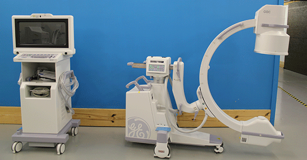
Our series continues for any of you who have ever been stumped by the acronym and tech-laden conversations of the radiology world. And, of course, no imaging glossary would be complete without a peek at common C-arm acronyms and phrases.
C-Arm Terms Glossary
CCD Camera
A CCD (charge-coupled device) camera is a camera which incorporates a CCD to create high-resolution images under a variety of light conditions.
To maintain your C-arm’s image quality, the CCD camera needs to be periodically cleaned and calibrated. In models without digital detectors, the CCD camera is coupled with another important C-arm part commonly called the II, which we'll get to in a minute.
Cine
This is a slang term, short for cinefluorography or cineradiography. Cine is, essentially, an X-ray “motion picture”. Using cine allows real-time viewing during procedures like needle placement or surgery.
CPU Board
One of many circuit boards in your C-arm, the central processing unit (CPU) is the main core and "brain" of your system's computer. A functioning CPU board allows the internal components of your C-arm to communicate and work in tandem.
DICOM
This is an acronym for a file type developed specifically for medical images. Digital Imaging and Communications in Medicine (DICOM) is the industry standard file type for handling, storing, printing and transmitting information in medical imaging. DICOM files can be exchanged between imaging systems, printers, and physician workstations that are DICOM-enabled.
DSA
Digital Subtraction Angiography, software feature that removes or “subtracts” bone or tissue from an image for clearer viewing of the vascular system.
FPS
Frames Per Second, how many images the cine camera can take in one second.
Gamma Correction
Gamma correction controls the overall brightness of an image. Images which are not properly corrected can look either bleached out, or too dark.
II
Image Intensifier, a light amplification device that converts x-ray photons to light photons, makes x-ray images bright enough and magnified enough for proper viewing
- 9” II ( tri mode 9”,6” 4”)- higher magnification than 12”
- 12” II (tri mode 12”, 9” 6”) – primarily used for vascular studies (larger field of view)
MARS
Motion Artifact Reduction System, software that reduces incidents of image distortion created by movements of the subject.
MTS
Motion Tolerant Subtraction, software that functions much like standard DSA but offers additional benefits that reduce the interference of motion in the subject during scanning, also allows for full run-offs in a single injection and run. (GE Healthcare 2012)
PACS
PACS: Picture Archiving and Communication System – A medical imaging network technology which provides economical storage of, and convenient access to, images from multiple modalities.
The PACS server plays an integral role in the exporting, sharing and storing of patient information and images. From larger cardiac cine sequences to individual spot films, this server can host a multitude of different studies from a plethora of modalities. Getting a PACS server set up and communicating properly can be a very tedious process. Do your homework and make sure that you’re bringing in a qualified PACS administrator to complete this operation.
If we left a phrase you hear every day off the list, please let us know! Help us make this blog series as useful as possible for anyone needing a quick reference point in the imaging community.

Josh Block
Josh Block is the President of Block Imaging. As a part of the second generation of Blocks to lead Block Imaging, Josh carries on the family passion for putting people first, delivering memorable service, and improving lives all over the world by empowering medical care providers with high-quality equipment. Aside from work Josh enjoys being a husband and dad, playing golf. And, before turning 40, Cheetos and cherry coke.


