If you keep your ear to the ground in the world of CT scanner technology, you've probably heard the name "Gemstone Spectral Imaging" (GSI) rolling around in conversations about the latest CT models from GE. You've probably also heard that GSI allows for better pictures of blood vessels, improved viewing of joint replacement, and more accurate diagnosis of numerous other health conditions1. But, while the name sounds impressive and all of those benefits are highly desirable, they don't go very far in explaining what GSI is and how it works.Today, we hope to clear those items up for you.
What Gemstone Spectral Imaging Is
GSI is GE's take on the spectral imaging concept, that is, using two separate energy sets to examine the different attenuation properties of matter2. In layman's terms, a CT system equipped with GSI, or another spectral imaging product, will take data sets at two levels of X-ray power in a single scan. The two data sets are then compared within the image reconstruction software and offer an additional level of detail.
How Gemstone Spectral Imaging Works
There are three components to the function of GSI: hardware, technique, and software.
Hardware
The real namesake of GSI is the hardware portion, the Gemstone detector. GE describes this new detector as having “jewel-like material properties". Its components synthetically replicate the structure of garnet crystals3 (think cubic zirconium- looks like a diamond, without the expense). This product is the classic transparent ceramic detector, evolved. The Gemstone scintillator escalates response to X-ray excitation, reportedly 50 to 100 times faster than any other detector on the market. The higher speed improves temporal and spatial resolution and reduces electronic noise.
Technique
As we touched on earlier, the main acquisition technique behind GSI is the use of dual energy CT (DECT). With this technique, power is quickly switched between two different energy levels, multiple times in a single rotation2, making it easier to characterize tissue and tease out metal artifacts (previously problematic in image analysis). The technique makes it easier to differentiate between features based on chemical composition and material density. For example, DECT can help discern arterial plaque during CT angiography (GSI Cardiac). It also allows the effect of contrast iodine to be virtually dialed up and down to improve images of blood vessels1 and allows more accurate perfusion calculations.
GSI has been helpful in identifying even small lesions in abdominal scans and can potentially determine the type of kidney stone a patient has, allowing the most appropriate remedy to be prescribed. Compared to typical PET, SPECT, and MRI procedures, the technology allows quicker, more comfortable scans of patients.
Software
After acquisition, the image data goes into the reconstruction software process. According to GE, "Projection-based reconstruction is used to process the data. Based on known attenuation curves, the process mathematically transforms low and high kVp attenuation measurements into effective material density (MD) basis-pair images."3 Essentially, GSI images have detail above and beyond standard CT based on the software's projection of how the anatomy in the shot resisted the flow of radiation at each power level.
Where Can Gemstone Spectral Imaging Be Found?
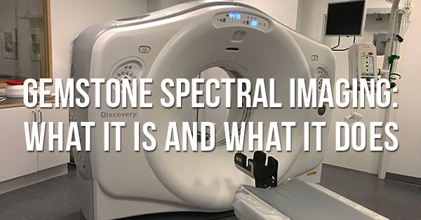
Gemstone detectors and Gemstone Spectral Imaging features can be found on GE's Revolution GSI, Revolution HD CT, and Discovery CT750 HD4. These are high-end systems. most of which won't begin finding their way to the secondary market for several more years.
If your needs have you looking to upgrade your CT sooner than later, but you feel you absolutely must have GSI, you'll probably have to purchase directly from GE. If your budget isn't ready to handle a brand-new purchase price, you'll need to wait for GSI systems to come off their leases and trickle onto the used market. In the meantime, there are other options from GE that can serve you well. Our team is ready to discuss those options with you to find a good fit while all of us wait for used, GSI-enabled scanners to become available.
1 https://www.insideradiology.com.au/dual-energy-ct-scan/2 https://radiopaedia.org/articles/dual-energy-ct-1?lang=us
3 https://www.itnonline.com/article/spectral-imaging-brings-new-light-ct

Paul Crawford
Paul Crawford is the Vice President of Equipment Solutions at Block Imaging. Paul connects with healthcare facilities across the world to offer CT solutions and manages the wholesale sales team. When Paul is not helping customers with their CT needs, he enjoys spending time with his family, watching MSU sports, and CrossFit.


